Definition
Causes and Influences
Epidemiology
Pathology
Clinical features
The joints
The skin
Cardiovascular
Pulmonary
Gastrointestinal
Renal / Kidney
Central Nervous System
Reticuloendothelial
The Laboratory investigations
Antibodies in SLE
Criteria for SLE
Assessment of severity
Principles of therapy
Definition - SYSTEMIC LUPUS ERYTHEMATOSUS - SLE / LUPUS
SLE is a chronic
inflammatory illness characterised by a immunoregulatory disturbance and
producing a multisystem disorder including skin, joints, blood, kidney,
lung, brain, and other organs.
It was originally described by BIETT in 1822.
The term LE was given by CAZANAVE in 1851
The systemic manifestations were described by
KAPOSI and OSLER
HARGRAVES first described an unusual cell
type on histology in these patients - the LE CELL. HASERICK associated
this LE CELL with ANTINUCLEAR ANTIBODIES - antibodies found present in
the lupus patients.
Various
influences are associated with the disease. These include:
GENETIC
HORMONAL
ENVIRONMENTAL
The prevalence is between 2.9-400 / 100000
The disease is much more common in females -
Female to male ratio is 9 : 1, and occurs also in greater numbers in
Blacks and Asians.
However , certain communities are found to have
high prevalence rates.
There is a high prevalence in the Western Cape
of South Africa - for example amongst persons of mixed ancestry, whilst
it is noted to be rare amongst the rural black population of South
Africa.
Definite FAMILIAL relationships are noted and
genetic profiles have been described.
HLA A1
B8 DR3
NULLC4
ALLELE
ACETYLATOR STATUS - a mechanism of enzyme
degradation of certain nitrogenous chemicals in the liver.
REDUCED C3 complement receptors on Red blood
cells
ENVIRONMENTAL factors are also found :
Sunlight.
Dietary.
Drugs : Especially : Hydrallazine, Procainamide,
INH, and Certain Anticonvulsants- hydantoins, Aldomet, Certain
anti-thyroid drugs, Quinidine, Penicillin and Sulphonamides These have
been noted to occasionaly cause a drug induced Lupus like syndrome.
(Note : This does NOT prevent their use in patients with SLE).
HORMONAL Influences are also considered
important in pathogenesis. These include especially estrogenic
influences.
Perhaps the major mechanism of disease however
is the Immunological abnormalities.
These are manifested in several ways
LYMPHOPENIA
REDUCED SUPPRESSOR FUNCTION
REDUCED PRODUCTION / RESPONSE TO IL2 REDUCED KILLER CELL ACTIVITY
REDUCED PRODUCTION IL1
INCREASED B CELL STIMULATING FACTORS
AUTOANTIBODY PRODUCTION
POLYCLONAL B CELL ACTIVATION
ANTI-CELLULAR ANTIBODY
PLATELETS
RBC
LYMPHOCYTES
MEMBRANE PHOSPHOLIPID
IMMUNE COMPLEX FORMATION / DEPOSITION
ABERRANT IMMUNOREGULATION
LYMPHOCYTIC INFILTRATES
PERIVASCULAR INFILTRATES
MUSCLE CELL DAMAGE
HAEMATOXYLIN BODIES AND INCLUSION BODIES OF LE
CELLS:
ANTIBODY TO CELLULAR NUCLEOPROTEIN.
GLOBULAR MASS OF BLUISH HOMOGENOUS MATERIAL ON
H&E STAIN
CONCENTRIC FIBROSIS “ONION SKIN LESIONS” IN THE
SPLEEN
VERROUCOUS ENDOCARDITIS LESIONS IN THE HEART
VALVES
It is a MULTIPLE ORGAN
DISEASE characterised by EXACERBATIONS AND REMISSIONS.
Joint and
Musculoskeletal manifestations
ARTHRALGIA - This is common and is pain in the
joints with little to find on examination -other than regional
tenderness. The pain can be severe and is often worse in the morning,
and associated with some stiffness-especially in the hands. However the
patient often complains of continuous pain ALL over.
ARTHRITIS - This is characterised by the
presence of swelling of the synovial joint lining and is SYMMETRICAL and
usually NON EROSIVE
JOINT DEFORMITY may be seen - although uncommon.
A variety of ulnar deviation which is reversible is seen in the hands
called Jaccouds arthropathy.
TENOSYNOVITIS - this is a tendonitis and is
often seen in particular in the flexor hand tendons, but may manifest
also as regional enthesitis or bursitis problems.
AVASCULAR NECROSIS is an orthopaedic problem
which is associated with the disease but also with use of medications in
the disease - especially STEROIDS
Clearly the most visible and common
manifestation of SLE is the skin disease :
This is characterised by inflammation at the
DERMAL EPIDERMAL JUNCTION with :
OEDEMA - swelling
VACUOLISATION
PERIVASCULAR INFILTRATES of inflammatory cells
FOLLICULAR PLUGGING - plugging and blocking of
the follicles
DERMAL ATROPHY - thinning of the dermal skin
layer
IMMUNOFLUORESCENCE GRANULAR IgG DEPOSITS -
immune antibody fixation in the skin and dermis layer.
The skin disease has several subtypes -
classified according to the severity and type of rash.
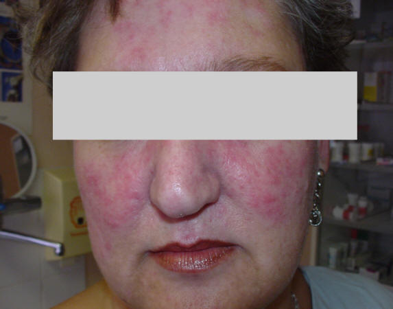
ACUTE CUTANEOUS L.E - This is characterised by
the classical BUTTERFLY RASH. It tends to EXACERBATE WITH FLARES of the
SLE and is characterised by
ERYTHEMA
OEDEMA
PHOTOSENSITIVITY
SUBACUTE CUTANEOUS L.E :
This is a rash seen in patients and is described
as SYMMETRICAL SUPERFICIAL NON SCARRING It is distributed especially on
the
NECK,
CHEST,
SHOULDERS,
BACK,
ARMS.
There are circular ANNULAR LESIONS NON SCARRING
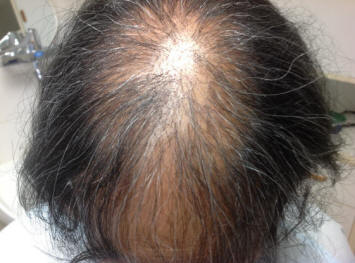
ALOPECIA - hair loss, which is patchy in the scalp is seen in 50% of
cases. PHOTOSENSITIVITY is VERY common finding.
An ASSOCIATION with certain genetic markers are
seen - HLAB8 DR3 ENA(Ro)
Interestingly , in this variety of SLE there is
a LOW INCIDENCE of LUPUS NEPHRITIS
It affects the SCALP EAR FACE NECK.
EARLY: changes seen include OEDEMA and the skin
is INFLAMED and HYPERPIGMENTED. LATER: The skin develops CENTRAL
DEPIGMENTATION and ATROPHY with DEPRESSED SCARS. TELANGIECTASIA -
prominent spidery like superficial blood vessels become prominent.
SEVERE ALOPECIA is common In discoid LE.
90% of patients have disease RESTRICTED TO THE
SKIN and there is a LOW INCIDENCE of SEVERE COMPLICATIONS and systemic
disease.
Positive antibody serology is usually seen but
is USUALLY a LOW TITRE
Cardiovascular manifestations:
PERICARDITIS - inflammation of the lining around
the heart is seen in 30% and presents with chest pain , malaise and
sometimes breathlessness.
The clinical findings may be absent with a
pericardial effusion identified on an XRAY or cardiac ultrasound..ie it
can be ASSYMPTOMATIC.
However a pericardial RUB may be heard on
auscultation of the heart on examination.
Occasionally the fluid in the pericardial space
can increase in volume, and rarely the fluid in the confined space can
compress the heart itself and result in CARDIAC TAMPONADE- with heart
failure. In addition, scarring down of the pericardium can result in
CARDIAC CONSTRICTION, another cause of right heart failure.
MYOCARDITIS occurs in 25% of patients and is
manifested by persistent TACHYCARDIA, ST-T CHANGES on electrocardiogram,
congestive heart failure with breathlessness, and ARRHYTHMIA with
palpitations or tachycardia complications.
ENDOCARDITIS implies involvement of heart valves
with inflammation and sterile VEGETATIONS called LIBMAN SACKS
VEGETATIONS.
The MITRAL and AORTIC valves are most commonly
involved.
The problem ranges from MILD to SEVERE.
There is an association between the valvular
lesions and the ANTI PHOSPHOLIPID ANTIBODY / Lupus anticoagulant.
EMBOLI are a potential complication of clots to
the brain and there is a potential for stroke. Involvement of BLOOD
VESSELS, may cause significant morbidity. This can be the result of a
vasculopathy or vasculitis, or due to accelerated atherosclerosis - a
potential complication of steroid therapy.
CORONARY ARTERITIS and STEROID related
ATHEROGENESIS may cause myocardial infarction.
PERIPHERAL VASCULOPATHY is also well documented.
There are several manifestations of this:
RAYNAUDS syndrome
LIVEDO RETICULARIS- a lace like rash on the
body, which is cold sensitive.
ANTIPHOSPHOLIPID ANTIBODY is seen more commonly
in patients with lupus and used to be called the lupus anticoagulant.
This is associated with, mid-trimester abortions, thrombosis of blood
vessels- arterial and venous, central nervous system disease and
headaches / migraine, mitral valve lesions and also thrombocytopenia -
low platelet counts.
PERIPHERAL VASCULITIS
PURPURA
URTICARIAL VASCULITIS
ANGIOEDEMA associated with C2 / C4 DEFICIENCY
and C1 ESTERASE INHIBITOR DEFICIENCY
LARGE VESSEL VASCULITIS - a process which is
relatively INFREQUENT
VASCULITIS of small and medium vessels may be
seen more commonly and LYMPHOCYTES/GRANULOCYTES are seen in the walls of
the vessels.
LEUKOCYTOCLASTIC CHANGE is more frequent with
RUPTURED CELLULAR DEBRIS.
IMMUNOGLOBULIN / C3 DEPOSITS may be identified
in the walls of the blood vessels.
PNEUMONITIS - inflammation within the lung
tissue - similar to pneumonia.
PULMONARY HAEMORRHAGE - pulmonary bleeding.
INTERSTITIAL LUNG DISEASE - a progressive
scarring of the lung tissue.
SHRINKING LUNG SYNDROME - an interesting feature
where the lung volumes decrease. This is felt to relate to elevation of
the diaphragms as a consequence of weakness of the muscular diaphragm.
INFECTIONS - with pneumonia and sinusitis must be distinguished by
disease activity. STEROIDS / IMMUNE THERAPY may reduce the host
defences, and infection may follow rapidly.
PULMONARY EMBOLISM - occurs with the development
of clot in the leg, which dislodges and travels to the lung vessels -
obstructing them. The result is chest pain and breathlessness. PULMONARY
HYPERTENSION. - With the occlusion and narrowing of the blood vessels,
and with the scarring and fibrosis of the lung tissue, as well as a
general increase in tone of the blood vessels within the lung, the
pulmonary pressures rise, resulting in breathlessness, and potential
progressive right sided heart failure - COR PULMONALE.
ESOPHAGEAL DYSMOTILITY - results in dysphagia -
difficulty swallowing food with a tendency for the food to get “stuck”.
There is an association with this and RAYNAUDS.
ABDOMINAL PAIN : One of the most interesting and
difficult management problems in SLE. This can be ACUTE/SUBACUTE and has
several mechanisms:
SEROSITIS - inflammation of the peritoneum - the
lining of the abdominal wall.
PANCREATITIS
VASCULITIS: which can result in visceral BOWEL
GANGRENE
VENOUS OCCLUSIONS
PEPTIC ULCERATION - especially related to DRUGS:
- the Nonsteroidal anti-inflammatories and high dose cortisone.
HEPATITIS - and inflammation of the lining
around the liver -peri-hepatitis, is an additional potential problem,
although generally uncommon.
Inflammation in the kidney is called nephritis.
This can be renal GLOMERULAR involvement or INTERSTITIAL involvement .
50% OF PATIENTS develop renal disease.
Several subtypes are known.
1. NORMAL
2. PURE MESANGIAL NEPHRITIS with MESANGIAL CELL
PROLIFERATION AND MATRIX THICKENING
3. FOCAL SEGMENTAL Glomerulonephritis (GN) -
with SEGMENTAL PROLIFERATION
4. DIFFUSE PROLIFERATIVE GN with PROLIFERATION
/CRESCENTS / and FIBRINOID MATERIAL visible in the glomeruli
5. MEMBRANOUS GN. - with BASEMENT MEMBRANE
THICKENING
6. ADVANCED SCLEROSING GN. - with END STAGE
FIBROSIS and scarring
Mortality from renal disease has improved
dramatically over the past few years with identification of the more
severe subtypes and appropriate therapy.
DIALYSIS has also offered patients with end
stage renal failure an opportunity for prolongation of life, and RENAL
TRANSPLANTATION has where available, given these patients a totally
improved quality of life, although immunosuppression is required, as
well as ongoing follow up in these patients. SURVIVAL in Lupus Nephritis
is now approximately.
85% over 5 YEARS and
65% over 10 YEARS.
Therefore an examination of the urine for blood
and protein is vital as well as an examination of the urine sediment
with a microscope. An increase in the protein or blood in the urine is a
possible indication for renal biopsy to determine the subtype of renal
disease and determination of aggression and type of therapy.
CNS - Central Nervous System disease
CNS disease Is extremely common and has a wide
spectrum of involvement.
DEPRESSION
PSYCHOSIS
EPILEPSY
HEADACHES
MIGRAINE
TIA - transient ischaemic attack with transient
weakness or neurological deficit.
STROKE - established neurological deficit.
CHOREA - an involuntary movement disorder
MYELOPATHY - spinal cord disease.
POLYNEUROPATHY - peripheral nerve disease - with
weakness or sensation disturbance in the limbs.
Reticuloendothelial involvement.
HEPATOMEGALY - an enlargement of the liver.
SPLENOMEGALY - an enlarged spleen
The laboratory investigations in SLE.
I generally do a Blood Count, ESR (Sedimentation
rate) and a CRP - C-Reactive Protein.
The CRP is usually not elevated in lupus unless
there is coincidental infection or inflammation from another cause. The
ESR is usually elevated in active disease or inflammation and infection.
HAEMOLYTIC ANAEMIA - due to antibody destruction
of Red blood cells as part of the autoimmune process is a potentially
serious problem and possibly requires Corticosteroids or more potent
immunosuppression for more severe involvement.
LEUCOPENIA - a low white cell count is frequent,
and in particular a low lymphocyte count fraction may be noted.
THROMBOCYTOPENIA - a low platelet count is seen
in 25% of patients.
This is either due to marrow suppression -
either from the disease or drugs used, especially the cytotoxic drugs,
or peripheral consumption with destruction of platelets in the
circulation.
A bone marrow examination is required to
distinguish which source the problem is from. The peripheral destruction
of the platelets is due to antibody against the platelets themselves
part of the autoimmune process.
I also then do a spectrum of antibody tests -
including, antinuclear antibody ANA, anti DNA antibody, extractable
nuclear antibodies - especially ENA sm, ENA rnp, and antihistone
antibody if drug induced lupus is suspected.
These are further discussed below.
A measurement of IMMUNE COMPLEXES may give an
assessment of the level of circulating antibody - antigen complexes
COMPLEMENT- These Immune molecules involved in
inflammation are consumed by these complexes and their levels drop, and
also can be measured as an index of disease activity.
A renal function assessment should include a
test for blood and protein and microscopy. A blood Urea nitrogen and
Creatinine, as well as a 24 hour urine collection to investigate the
Creatinine clearance and protein production over 24 hours is necessary.
Where appropriate, a liver function is done.
ANA
DS DNA (Double stranded DNA)
ENA
ANTI HISTONE antibody.
The Antinuclear antibodies :
An antibody is a protein made by the B cell
lymphocyte of the body, under the stimulus of the T lymphocyte cell. The
initial T cell response follows presentation to the T Cell of antigen,
by an “antigen presenting cell” - usually a macrophage.
The original stimulating antigen in lupus is not
known, but felt by many to be an infective agent - possibly a virus or
retroviral particle.
The T Cell requires a particular “self” gene
marker to respond to the cell presenting the antigen to it. If it does
not see these self gene markers - no reaction will occur.
The “self” gene marker is related to the HLA
gene series - that provides markers for risk factor of disease.
The antibodies in Lupus are associations with
the disease and not necessarily causal.
Thus damage to an organ may expose tissue
antigens to the macrophage, and antibodies may therefore be produced as
a result of damage, rather than causing the damage in the first place.
These antibodies are antibodies to “self” and
hence the terminology - autoimmune - “immune to self”
The tests done are :
Chronic active hepatitis
Primary Biliary cirrhosis
Pulmonary fibrosis
Sjogren syndrome
Drug induced lupus
Bacterial endocarditis
Old age
This is a FLAGELLATE WITH A KINETOPLAST
CONTAINING HELICAL double stranded DNA WITHOUT HISTONES.
Antibody can be made to detect this.
DNA BOUND TO KINETOPLAST IS DETECTED BY
FLUORESCENT ANTIGLOBULIN SERUM.
These antibodies are directed against RNA
EXTRACTED FROM THE NUCLEUS WITH SALINE.
They are LABELLED WITH PHOSPHOROUS AND
PRECIPITATED WITH ANTIBODY.
Several are described:
ANTI -U1 RNP Other than SLE may be seen in
Mixed connective tissue disorder :MCTD
MAY IDENTIFY PRECURSORS TO SYSTEMIC SCLEROSIS
They also identify a subset of lupus patients
with LESS RENAL DISEASE
ANTI-Ro (SS-A) Seen in:
SLE
SJOGREN syndrome
Associated with increased PHOTOSENSITIVITY in
SLE patients
Identify risk of NEONATAL HEART BLOCK in babies
of SLE patients
ANTI-Sm Seen in SLE
The Fluorescent Staining characteristics of the
antibodies are as follows
HOMOGENOUS / DIFFUSE STAIN
PERIPHERAL / RIM STAIN
Ab TO RNA :
NUCLEOLAR PATTERN
Ab TO EXTRACTABLE
ANTIGEN :
SPECKLED PATTERN
IN FACT : There are multiple clinical
manifestations and therefore Criteria are identified for diagnosis by
the American College of Rheumatology (ACR) These criteria are more for
the classification of the disease and do not mean that therapy must be
withheld because not all criteria have been fulfilled.
However they ensure that patients entered on
studies are definite cases and improve the validity of trials.
They also, of course, increase diagnostic
certainty, if all criteria are fulfilled.
CRITERIA FOR DIAGNOSIS SLE : Revised Criteria
For Classification (Require 4/11)
2. DISCOID RASH: KERATOSIS PLUGGING ATROPHY
3. PHOTOSENSITIVITY
BY PATIENT HISTORY OR CLINICAL
OBSERVATION.
4. ORAL ULCERS
USUALLY PAINLESS
5. ARTHRITIS
NONEROSIVE >1 JOINTS
6. SEROSITIS
PLEURA/PERICARDIAL
7. RENAL
PROTEIN >3+ / >0.5g CELLULAR CASTES
8. NEUROLOGICAL
SEIZURE PSYCHOSIS
9. HAEMATOLOGICAL HAEMOLYTIC ANAEMIA OR
LEUKOPENIA OR LYMPHOPENIA OR THROMBOCYTOPENIA
10. IMMUNOLOGICAL
+LE CELL PREPARATION OR ANTI DNA OR SM
ANTIBODY OR FALSE +VE SYPHILIS
11. ANTINUCLEAR ANTIBODY
BY IMMUNOFLUORESCENCE OR EQUIVALENT
ASSAY
Once the diagnosis is made , it is important to
evaluate the severity of disease and establish the degree of organ
involvement This is done clinically and with laboratory tests.
I still believe that many aspects of Lupus
therapy is also an art rather than only a science.
The distinguishing of activity flare from
infection for example is often extremely difficult, as often a source of
infection is not identifiable, and Lupus patients are more susceptible
to severe infections. Indeed - this is where the experience of the
Rheumatologist or Physician is so critical - and therein lies the ART of
lupus therapy. Of course the lab and science is equally important and
many new advances are discovered with research.
Assessment of disease Severity
CLINICAL
The presence
of severe ORGAN INVOLVEMENT Especially :
NEPHRITIS
CVS
SKIN
SEROSITIS
FEVER /
FATIGUE / WEIGHT LOSS
High levels
of antibodies ANF / ANTI DNA
LOW levels
of COMPLEMENT
High levels
of immune complexes
SLE :
The Principles of Therapy
Once an assessment is made of disease activity -
a plan can be made for therapy. Always remember that Lupus patients also
get all the common minor illnesses seen with non-lupus individuals. Not
every problem is due to the Lupus, and not every problem represents a
complication thereof.
There is a tendency for some Doctors and Medical
personel to fear the disease, which is a problem that is lessened with
the experience of the practitioner.
It is however very important to remain vigilant.
Therefore it is recommended that a
Rheumatologist see the patients and hopefully coordinate therapy, but
involve the Family physician, and other paramedical staff - including
Physical therapists, Occupational therapists, other involved staff and
certainly, the patient and family concerned.
EDUCATION is very important
2. MANAGEMENT of the CHRONIC DISEASE
For the joint manifestations - judicial use of
Nonsteroidal anti-inflammatories (NSAIDS) are almost always required -
especially if there is swelling and stiffness of the joints. Analgesics
are useful for pain control - and should be used depending on severity
of the discomfort. Antimalarials ie plaquenil, are particularly useful
for joint and skin disease and should be seen as a controller of the
underlying process, rather than symptomatic therapy alone. For the skin
disease, topical cortisone preparations in different strengths are
useful. For the face, topical steroids should be avoided unless in
dilute format as they can cause skin thinning. However severe disease
sometimes warrants usage under guidance of a dermatologist if necessary.
Antimalarials are useful for skin disease control. Monitoring of eye
toxicity is required for long term use of the antimalarials.
Use of corticosteroid tablets should be used for
severe disease, where function is impaired and NSAIDS are not helping
for joint problems, and for major organ involvement. Corticosteroids can
be used in oral form or in intravenous “pulse therapy” for severe organ
disease.
Examples include
CYCLOPHOSPHAMIDE
AZATHIOPRINE
METHOTREXATE
mycophenolate
Therapeutic measures sometimes employed include
Dapsone
PLASMAPHORESIS
TOTAL LYMPHOID IRRADIATION
Biologic drugs
Rituximab anti cd20 antiboby given as 500mg x 2
iv infusions 6 monthly
DIALYSIS has altered the prognosis of millions
of patients in end stage renal failure, as has transplantation for
thousands of individuals.
For chronic disease the aim of therapy is :
PERIODIC EVALUATION
PREVENT ORGAN DETERIORATION
MINIMISE DISABILITY
MINIMISE COMPLICATIONS OF THERAPY
It is important to establish a maintenance
disease modifying therapy regimen if possible - including antimalarials
and if severe disease, as minimal doses of NSAIDS, analgesics, cortisone
and immunosuppression drugs as the cost - benefit side effect profile
ratio will allow.
| Involvement | Symptoms |
| Constitutional symptoms | Fatigue, brain fog and fevers |
| Alopecia | Hair loss |
| Skin rashes |
-An acute classical butterfly distribution rash on the face with
sensitivity to light.
-A subacute non-scarring skin rash on the neck, chest and
shoulders.
-A chronic scarring type called discoid lupus with scarring
of the skin.
|
| Mouth ulcers | Usually painless, but may increase as the disease progresses |
| Joint manifestations |
Arthralgia: morning stiffness, pain without swelling
Arthritis: swelling in the joints
|
| Cardiac involvement |
Endocarditis: inflammation of the heart valves
Myocarditis: inflammation of the heart muscle
Pericarditis: inflammation of the lining of the heart
Varying severity of chest pain or heart failure with
breathlessness
|
| Respiratory involvement |
Pleurisy: inflammation of the lining of the lung with chest pain
on breathing, or discomfort with breathlessness caused by damage
to lung tissue |
| Vascular involvement |
Raynaud’s: white, blue and red colour changes in the fingers in
the cold.
Livedo: lacy pattern of skin pink / blue colour. Also seen with
temperature changes.
Vasculitis: severe inflammation of the blood vessels to the skin
or internal organs.
Hypertension is common, aggravated by kidney disease.
|
| Blood disorders | Marrow involvement, or autoimmune peripheral blood involvement, with moderate to severe anaemia or bleeding disorder because of platelet reduction or low white count and increased infection risks |
| Clotting disorders | Antiphospholipid syndromes, which may cause thrombosis of vessels or embolism to the lung. |
| Gastroenterological involvement | Swallowing issues, abdominal pain or, rarely, pancreatitis or hepatitis. |
| Kidney disease | Varying severity of inflammation of kidney tissue causing blood or protein in urine on testing. This is the most important involvement in lupus, and in the past was the primary cause of death in patients from kidney failure. |
| Pregnancy issues | Fertility and second trimester foetal loss is associated with lupus, especially if antiphospholipid antibodies are found. Hypertension and aggravated kidney disease and bleeding and clotting issues with pregnancy cause potential harm to mother and child. It is essential to control the disease before pregnancy can be undertaken. |
Dr Gotlieb
Rheumatologist.