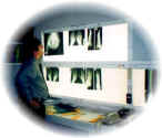
The radiology department
The radiology department is that part of the hospital, where images of the joints are obtained by different techniques, all of which are designed to visualise different tissues. Such methods, commonly used in rheumatology, include:
Plain radiography - X-rays
Scanning - Magnetic resonance MRI and CAT scanning (computerized
tomography)
Radio-labeled isotope scanning (Often done in the nuclear medicine
department)
Bone density scanning to assess osteoporosis.
Radiography is not only restricted to the joints, but also includes radiology of heart and lungs and brain. This is because arthritis diseases often involve internal organs.
Radiology, like the laboratory, is often overemphasised in the diagnosis of rheumatic disease, as many diseases that present early, have normal x-rays. Therefore it is mistaken, that just because the x-rays are normal, that there can be no problem. In fact, we as rheumatologists, actually want to see normal X-rays. The absence of damage is desirable, and it is these patients, especially, who should get appropriate therapy to prevent damage.
The radiologists therefore form an adjunct to the clinician in reaching a clinical diagnosis.
X-rays reflect a historical record over ones lifetime, of joint damage or change, with poor reflection on the current activity of disease. Conventional X-rays are done by projecting a beam of x-rays through the body region and onto a photographic plate. The X-rays penetrate different tissues differently, and therefore we see a good image of the skeletal structure and a much poorer, variable image, of the surrounding soft tissue. The investigation has some problems of reproducibility, as pictures vary with the technique of the radiographer. They are used to establish baseline status, as well as progression and therapeutic response over time.
When we look at a basic X-ray of a joint, we look at the soft tissue shadows around the joint, but also look at the joint space which represents the state of the cartilage. Damage to the cartilage appears as a narrowing of the joint space. We then look at the bone margins - looking for erosions or holes in the bone surface within the joint. Erosions represent damage and are seen in arthritis groups characterized by inflammation. We then look at the bone itself. Thickening of the bone is called sclerosis and is seen in osteoarthritis with degenerative disease of the joint This will occur especially on the weight bearing side of the joint. In addition, bony outgrowths, called osteophytes are seen at the edge of the bone margins and are called osteophytes. In addition Thinning of bone is called osteopenia or osteoporosis, and is seen in inflammatory arthritis - especially rheumatoid arthritis, due to increased blood flow from the inflammatory process. Generalised osteoporosis requires more accurate testing and X-rays in isolation are a poor measure of bone loss itself.
Scanning may be done using X-rays, as in CAT scanning (computerised axial tomography) or magnetic resonance imaging - MRI. The former is done by taking multiple X-ray slices, then assimilating these to form a picture. It is useful in visualising internal organs, but has largely been superceded by MRI. MRI requires a person to be placed in a long and narrow tunnel, and to have a magnetic field initiated and terminated within the tunnel, and then measure release of energy released from this cycling magnetic field. The patient has to remove all metal magnetic objects and should expect to be slightly cramped in space. The tunnel may be noisy one the scan commences. The duration of the scan depends on the part being scanned. The patient is required to lie still for approximately 10 - 15 minutes. However the pictures that are generated reveal fine detail of internal organs, and the scan is most useful in detecting soft tissue problems., as well as spinal and brain problems, organ anatomy and bone and joint detail.
Isotope scanning is a functional scan, that is used to determine uptake of radioactive material to a tissue site. There are several applications in rheumatology, but the commonest is Technetium scanning for detection of inflammation. The technique is to inject a material and then to allow it to circulate in the bloodstream, followed by scan 2 hours later, in which a scanning device picks up the trace nuclear medical material, and this is computer assimilated to form a image of the entire human or affected part in a picture not dissimilar in appearance to the Turin shroud.
The entire test takes 2 - 2.5 hours, with no claustrophobia, and is best indicated in early inflammatory arthritis. However it can also be very effective to highlight bone disease, microfracture, sepsis, tumors amongst other cause of disease.
Angiography is a technique where a dye is introduced into a vein or artery then take X-rays rapidly to look at flow through a small or narrowed artery.
Bone densitometry, is a technique to measure the density of bone, and also to calculate risk of fracture as a result of osteoporosis. The test is simple and convention has made the most common variety, the DEXA - Dual photon X-ray absorptiometry. The other main techniques are the use of QCT scan - quantitative CT scanning, and ultrasound of the heel.
The methods each have their individual strengths and weaknesses.
The X-ray department will be very useful to your doctor, but again you must remember that even in serious disease, the X-rays may be completely normal, and therefore the clinical examination remains the cornerstone of rheumatology
Remember - X-rays can be completely normal